human anatomy and physiology pdf notes
Ace your anatomy & physiology class with our concise, easy-to-understand PDF notes! Packed with visuals and key concepts, these notes are your secret weapon for success. Download now and conquer human biology!
Human Anatomy and Physiology PDF Notes⁚ A Comprehensive Overview
This comprehensive guide offers a detailed exploration of human anatomy and physiology. It covers the structural organization of the human body, from cells to organ systems, and their functions. The notes delve into key systems, including cardiovascular and nervous systems, providing essential information for students and professionals alike. Access readily available online resources to enhance your understanding.
Human anatomy and physiology is a fascinating field exploring the intricate structure and function of the human body. Anatomy focuses on the body’s physical structure, from its microscopic components like cells and tissues to its macroscopic organs and systems. Physiology, conversely, delves into how these structures work together to maintain life. Understanding the interplay between structure and function is paramount. This introductory section lays the groundwork for understanding the complexity of the human body. We will examine the organizational levels, from the simplest chemical building blocks to complex organ systems. The study of human anatomy and physiology is not merely an academic pursuit; it is crucial for advancements in medicine, healthcare, and our overall understanding of life itself. Many online resources offer supplementary information, including interactive diagrams, videos, and practice quizzes.
Levels of Organization in the Human Body
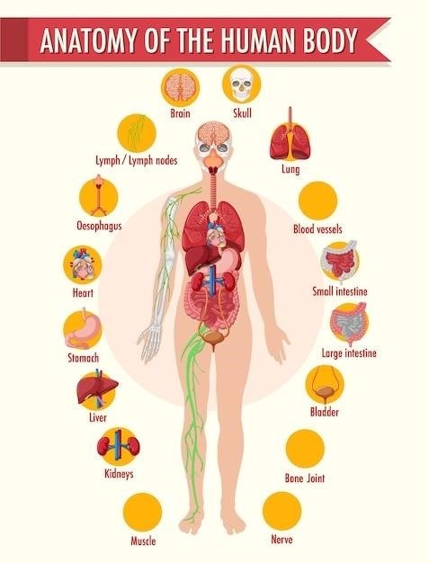
The human body exhibits a remarkable hierarchical organization, progressing from the simplest to the most complex levels. At the most fundamental level are chemicals, including atoms and molecules, forming the building blocks of life. These chemicals organize into cells, the basic structural and functional units of the body. Cells then aggregate to form tissues, groups of similar cells working together. Different tissues combine to create organs, which perform specific functions. Finally, multiple organs collaborate within organ systems, such as the cardiovascular or nervous systems, to maintain overall body homeostasis. Understanding these levels is crucial for comprehending how the human body functions as an integrated whole. Each level’s structure directly influences its function, and disruptions at any level can have cascading effects throughout the entire system. Detailed illustrations and diagrams are invaluable resources for grasping this hierarchical structure effectively. Explore various online platforms for visual aids and interactive learning tools.
Cells⁚ The Basic Units of Life
Cells, the fundamental building blocks of all living organisms, are microscopic structures exhibiting incredible complexity. Human cells, like those of other eukaryotes, possess a membrane-bound nucleus containing genetic material (DNA). This DNA directs the cell’s activities and determines its characteristics. Surrounding the nucleus is the cytoplasm, a gel-like substance containing various organelles, each with specialized functions. Mitochondria, often called the “powerhouses” of the cell, generate energy through cellular respiration. Ribosomes synthesize proteins, essential for cellular structure and function. The endoplasmic reticulum and Golgi apparatus modify and transport proteins, while lysosomes break down waste materials. The cell membrane regulates the passage of substances into and out of the cell, maintaining internal balance. Understanding the structure and function of these organelles is paramount to comprehending cellular processes and their roles in overall human physiology. Consult detailed diagrams and online resources to enhance your understanding of cellular components and their intricate interactions.
Tissues⁚ Groups of Cells with Similar Functions
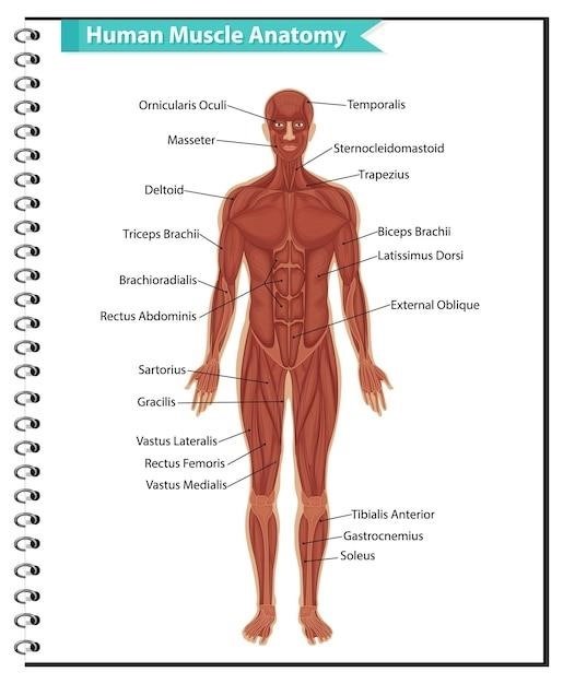
Tissues represent the next level of organization in the human body, arising from the aggregation of similar cells working together to perform specific functions. Four primary tissue types define the structural foundation of our bodies⁚ epithelial, connective, muscle, and nervous tissues. Epithelial tissues form coverings and linings, protecting internal organs and regulating substance transport across surfaces. Connective tissues, encompassing bone, cartilage, and blood, provide support, connect tissues, and transport substances throughout the body. Muscle tissues, including skeletal, smooth, and cardiac muscle, enable movement through contraction. Finally, nervous tissues, composed of neurons and glial cells, facilitate rapid communication and coordination within the body. Each tissue type exhibits unique structural and functional properties reflecting the specialized roles of its constituent cells. Understanding the characteristics of these four tissue types is crucial for grasping the organization and function of organs and organ systems. Further exploration of histological images and online resources will significantly deepen your understanding.
Organs and Organ Systems⁚ Complex Structures and Their Interactions
Building upon the foundation of tissues, organs emerge as intricate structures composed of two or more tissue types working in concert to perform specific functions. Examples include the heart, lungs, and kidneys, each exhibiting a unique architecture reflecting its specialized role. Organ systems represent the highest level of organization in the human body, integrating multiple organs to accomplish complex physiological processes. Consider the cardiovascular system, a network of the heart, blood vessels, and blood, responsible for transporting oxygen and nutrients throughout the body. Similarly, the digestive system, comprising organs from the mouth to the anus, processes ingested food for nutrient absorption. Understanding organ and organ system interactions is key to comprehending the integrated functioning of the human body. The seamless collaboration between these systems ensures the maintenance of homeostasis, a dynamic equilibrium essential for life. Effective learning resources, including online anatomical atlases and interactive simulations, enhance this understanding.

The Cardiovascular System
This section explores the intricate network responsible for circulating blood, delivering oxygen and nutrients, and removing waste products. Detailed study includes the heart’s structure and function, blood vessels, and the mechanisms regulating blood pressure. Understanding this system is vital for comprehending overall human health.
Anatomy of the Heart
The heart, a remarkable muscular organ, is the core of the cardiovascular system. Its four chambers—two atria and two ventricles—work in a coordinated sequence to pump blood throughout the body. The right atrium receives deoxygenated blood from the body, passing it to the right ventricle, which then pumps it to the lungs for oxygenation. Oxygenated blood from the lungs enters the left atrium, flowing into the left ventricle, the powerhouse responsible for pumping oxygen-rich blood to the rest of the body via the aorta. Valves, strategically positioned between chambers, ensure unidirectional blood flow, preventing backflow. The heart’s intricate structure, including its specialized muscle tissue (myocardium), conduction system (initiating and coordinating heartbeats), and coronary arteries (supplying the heart itself with oxygenated blood), is crucial to its efficient function. Understanding this detailed anatomy is fundamental to comprehending the heart’s role in maintaining overall circulatory health. The heart’s structure is beautifully complex, and its function is essential to life itself. A deeper understanding is revealed through detailed anatomical diagrams and histological studies. Detailed study of the heart’s chambers, valves, and specialized conduction system reveals the intricate mechanisms that ensure its rhythmic contractions and efficient blood pumping capabilities.
Blood Vessels and Circulation
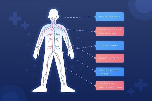
The circulatory system, a vast network of blood vessels, facilitates the continuous flow of blood, carrying oxygen, nutrients, hormones, and waste products throughout the body. Arteries, strong, elastic vessels, carry oxygenated blood away from the heart; their thick walls withstand the high pressure of blood ejected from the ventricles. Arterioles, smaller branches of arteries, regulate blood flow into capillaries. Capillaries, microscopic vessels with thin walls, allow for the exchange of substances between blood and tissues. Venules, small vessels collecting blood from capillaries, merge to form veins. Veins, thinner-walled than arteries, return deoxygenated blood to the heart; valves within veins prevent backflow, aided by skeletal muscle contractions. The circulatory system is divided into pulmonary circulation (heart to lungs and back) and systemic circulation (heart to the rest of the body and back). Understanding the structure and function of blood vessels, along with the mechanics of blood flow and pressure regulation, is critical to understanding cardiovascular health and disease. Detailed study of these vessels, including their microscopic anatomy and their role in nutrient and waste exchange, provides a more complete understanding of the circulatory system’s functions. The intricate interplay between different vessel types and the mechanisms that regulate blood flow are essential aspects of cardiovascular physiology.
Cardiac Cycle and Heart Sounds
The cardiac cycle represents the rhythmic sequence of events in a single heartbeat, encompassing contraction (systole) and relaxation (diastole) of the heart chambers. It begins with atrial systole, where the atria contract, forcing blood into the ventricles. This is followed by ventricular systole, where the powerful contraction of the ventricles ejects blood into the pulmonary artery and aorta. Finally, diastole allows the chambers to relax and refill with blood. These phases generate characteristic heart sounds, audible using a stethoscope. The “lub” sound (S1) is produced by the closure of the atrioventricular valves (mitral and tricuspid) at the start of ventricular systole. The “dub” sound (S2) is generated by the closure of the semilunar valves (aortic and pulmonic) at the end of ventricular systole. Additional sounds, such as S3 and S4, can sometimes be heard and may indicate underlying cardiac abnormalities. Understanding the precise timing and pressure changes during each phase of the cardiac cycle, coupled with the correlation of these events to the heart sounds, is crucial for diagnosing various cardiovascular conditions. The integration of electrocardiogram (ECG) data with the auscultation of heart sounds provides a comprehensive assessment of cardiac function.
Blood Pressure Regulation
Blood pressure, the force exerted by blood against vessel walls, is tightly regulated to ensure adequate tissue perfusion while preventing vascular damage. This intricate control involves a complex interplay of neural, hormonal, and renal mechanisms. Baroreceptors, pressure-sensitive sensors in the aorta and carotid arteries, detect changes in blood pressure and send signals to the brainstem’s cardiovascular center. This center adjusts sympathetic and parasympathetic nervous system activity to modify heart rate, contractility, and peripheral vascular resistance. The renin-angiotensin-aldosterone system (RAAS) plays a pivotal role in long-term blood pressure regulation. Reduced blood flow to the kidneys triggers renin release, leading to angiotensin II production, a potent vasoconstrictor. Angiotensin II also stimulates aldosterone secretion, promoting sodium and water retention, increasing blood volume and pressure. Antidiuretic hormone (ADH) from the posterior pituitary further contributes to water retention, while atrial natriuretic peptide (ANP) from the atria promotes sodium and water excretion, counteracting high blood pressure. These integrated mechanisms maintain blood pressure within a narrow range, adapting to various physiological demands and maintaining homeostasis.

The Nervous System
The nervous system, a complex network of specialized cells, governs communication throughout the body. It comprises the central nervous system (brain and spinal cord) and the peripheral nervous system, which includes sensory and motor neurons. This intricate system enables rapid responses to stimuli, coordinating bodily functions and higher cognitive processes.
Structure and Function of the Nervous System
The human nervous system is a marvel of biological engineering, a complex communication network responsible for virtually every aspect of our being. Its primary function is to receive, process, and transmit information, enabling rapid responses to internal and external stimuli. This intricate system is broadly divided into two main parts⁚ the central nervous system (CNS) and the peripheral nervous system (PNS). The CNS, composed of the brain and spinal cord, acts as the central processing unit, integrating sensory input and initiating motor output. The PNS, a vast network of nerves extending throughout the body, serves as the communication link between the CNS and the rest of the body. It carries sensory information from receptors to the CNS and transmits motor commands from the CNS to muscles and glands. Within the PNS, we find the somatic nervous system, which controls voluntary movements, and the autonomic nervous system, regulating involuntary functions like heart rate and digestion. The autonomic nervous system further branches into the sympathetic and parasympathetic systems, working in opposition to maintain homeostasis. The intricate interplay between these components allows for the seamless coordination of bodily functions, from basic reflexes to complex cognitive processes, highlighting the remarkable complexity and adaptability of the human nervous system.
The Brain and Spinal Cord
The brain, the command center of the central nervous system (CNS), is a remarkably complex organ responsible for higher-level functions like thought, memory, and emotion. It’s divided into three major parts⁚ the cerebrum, cerebellum, and brainstem. The cerebrum, the largest part, is responsible for conscious thought, voluntary movement, and sensory perception. Its convoluted surface, the cerebral cortex, houses billions of neurons, enabling sophisticated cognitive abilities. The cerebellum, located beneath the cerebrum, plays a crucial role in coordinating movement, balance, and posture. The brainstem, connecting the cerebrum and cerebellum to the spinal cord, controls essential life functions such as breathing, heart rate, and blood pressure; The spinal cord, a long, cylindrical structure extending from the brainstem, serves as the primary communication pathway between the brain and the rest of the body. It transmits sensory information from the periphery to the brain and relays motor commands from the brain to muscles and glands. The spinal cord’s segmented structure allows for localized reflexes, enabling rapid responses to stimuli without direct brain involvement. Protection of these vital structures is paramount; the brain is encased within the skull, while the spinal cord is protected by the vertebral column and its surrounding meninges. Understanding the intricate anatomy and physiology of the brain and spinal cord is essential to comprehending the complexities of the human nervous system;
Peripheral Nervous System
The peripheral nervous system (PNS) acts as the communication network connecting the central nervous system (CNS) to the rest of the body. Unlike the CNS, which is encased in bone, the PNS is more exposed, making it vulnerable to injury. It’s broadly categorized into the somatic and autonomic nervous systems. The somatic nervous system controls voluntary movements of skeletal muscles, allowing conscious control over actions like walking and talking. Sensory information from the skin, muscles, and joints is also transmitted via this system, providing awareness of the external environment. In contrast, the autonomic nervous system regulates involuntary functions, such as heart rate, digestion, and respiration. This system is further divided into the sympathetic and parasympathetic branches, which often have opposing effects. The sympathetic nervous system prepares the body for “fight-or-flight” responses, increasing heart rate and blood pressure. The parasympathetic nervous system, on the other hand, promotes “rest-and-digest” activities, slowing heart rate and stimulating digestion. Cranial nerves, originating from the brainstem, and spinal nerves, emerging from the spinal cord, form the major components of the PNS, extending throughout the body to innervate various organs and tissues. Understanding the PNS is crucial for comprehending how the CNS interacts with the body’s periphery, enabling coordinated function and response to stimuli.